Figure 6.
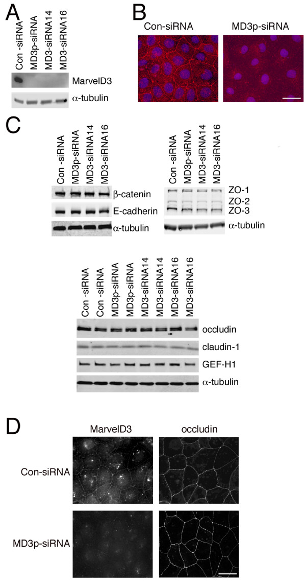
Depletion of MarvelD3 in Caco-2 cells. Caco-2 cells were transfected with the indicated siRNAs and then processed for immunoblotting (A and C) or immunofluorescence (B and D). (A, C) Cells were immunoblotted with antibodies against MarvelD3 and α-tubulin (A) or against a selection of tight and adherens junction proteins as indicated. Note, the expression levels of none of the junctional proteins apart from MarvelD3 were affected by depletion of the latter protein independent of whether filter- or glass-grown cells were analysed. Bar, 10 μm. (C) The lower panel in C shows duplicate cell extracts for each type of siRNA transfection. (B) Immunofluorescence staining of cells labelled with anti-MarvelD3 antibodies and Hoechst dye to stain DNA. Note, reduced levels of MarvelD3 were seen following transfection with MarvelD3 siRNAs. (D) siRNA transfected cells were labelled with anti-MavelD3 and anti-occludin antibodies. Bar, 10 μm. Note, knockdown of MarvelD3 did not appear to affect occludin distribution.

