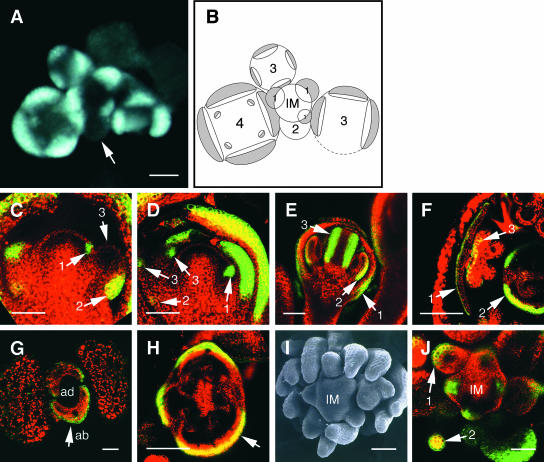
Figure 4.
Expression Analysis of FIL at the Reproductive Stage and in the fil Mutant.
(A) and (B) Transverse section of an fp-6 transgenic plant (see Figure 2A) examined by fluorescence microscopy (A) and its schematic view (B). Numbers in (B) indicate the stage of the floral buds. The GFP signal was observed in cells on the abaxial side of young flower primordia at stage 1. The signal was absent in the stage-2 flower primordium (arrow). In stage-3 and -4 flowers, the GFP signal was detected on the abaxial side of the newly formed primordia of sepals and petals. FIL-expressing regions are shown in gray.
(C) Arrow 1 indicates the GFP signal in the stage-0 primordium. In the stage-2 primordium, the signal was detected on the abaxial side of the basal region (arrow 2) but not in the upper region of the primordium (arrow 3).
(D) Longitudinal section of an inflorescence meristem. Arrow 1 shows the GFP signal on the abaxial side of a stage-1 flower primordium. Weak GFP expression remained on the abaxial side of the basal region of a stage-3 flower (arrow 2). GFP expression was induced on the abaxial side of sepal primordia (arrows 3).
(E) Longitudinal section of a stage-8 flower showing abaxial GFP expression detected in a sepal (arrow 1), a stamen (arrow 2), and a carpel (arrow 3).
(F) In a transverse section of a stage-12 flower, the GFP signal was observed in the petal epidermis on the abaxial side (arrow 1), on the abaxial side of the carpel (arrow 2), and on the abaxial side of the anther (arrow 3).
(G) Transverse section of a transgenic plant carrying fp-6 in the fil mutant. GFP was expressed on the abaxial side of the rosette leaves (arrow). The level and pattern of GFP expression in the fil mutant were the same as those in the wild type.
(H) Transverse section of a type-A flower of the fil mutant. Three sepals were formed in this flower, and the GFP signal was detected on the abaxial side of each (arrow).
(I) Top view of an inflorescence meristem of a fil plant showing a cluster of type-B filaments.
(J) Transverse section of a fil plant at the inflorescence meristem forming a cluster of type-B filaments. The GFP signal was observed on the abaxial side at the bottom of a filament (arrow 1) and on both the abaxial and adaxial sides in the upper region (arrow 2).
ab, abaxial side; ad, adaxial side; IM, inflorescence meristem. Bars = 20 μm for (C) to (F), (H), (I), and (J) and 50 μm for (A) and (G).

