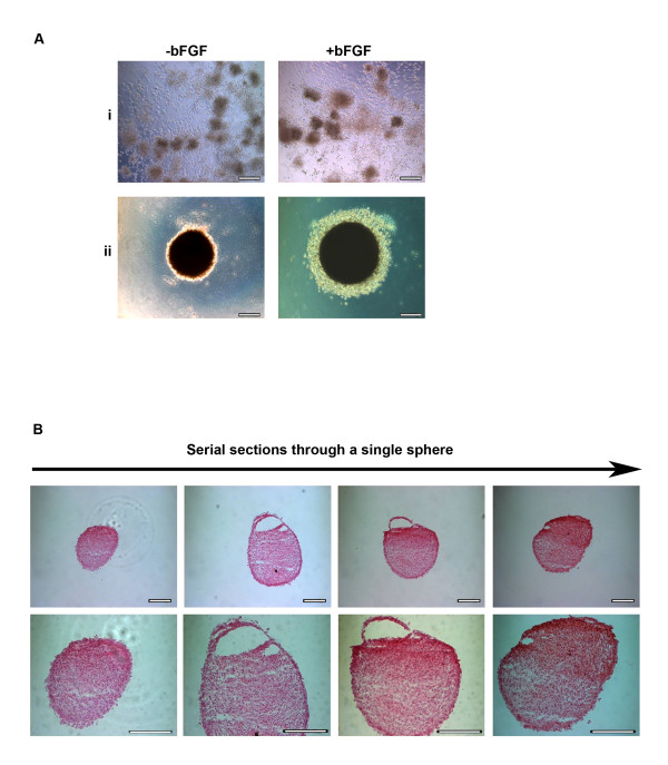
Figure 2.
Characterisation of spheres. A, Morphology of spheres seven days after seeding at high density (1 × 10⁴ cells/30 μl) as hanging drops in the presence or absence of bFGF in: i, stem cell proliferation medium without fetal calf serum; or ii, USSC growth mediumACF. (Magnification, ×200; scale bar is 100 μm) B, Serial sections of spheres stained with haematoxylin and eosin, generated by culturing in USSC growth mediumACF for seven days at both ×200 and ×400 magnification; scale bar is 100 μm.

