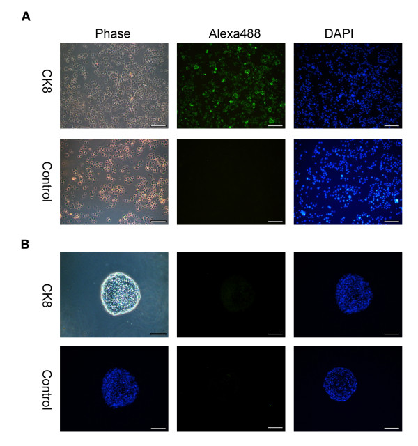
Figure 5.
Cytokeratin 8 staining on USSC-derived spheres. A, As a positive control for CK8 expression, primary human bronchial epithelial (16HBE14o-) cells were stained. B, Representative microscopy images of seven day spheres fixed, sectioned and stained with a Cytokeratin 8 (CK8) antibody (Alexa488; green) and counter stained with 4, 6-diamidino-2-phenylindole (DAPI; blue). The respective negative isotype control images are also shown. (Magnification ×200; scale bar is 100 μm.)

