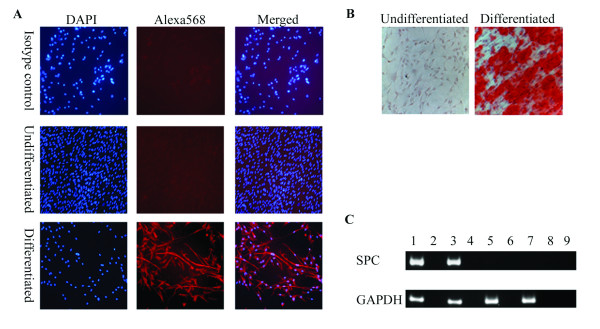
Figure 7.
Differentiation of spheres into neuronal, bone and epithelial-like cells. Seven days after sphere formation, cells were dissociated and plated on coated plates, as described in the materials and methods. The next day, cells were cultured in differentiation medium. A, Neuronal differentiation was confirmed by staining with a neuronal-specific β-tubulin III antibody (Alexa568; red) and counter stained with 4, 6-diamidino-2-phenylindole (DAPI; blue). B, Bone differentiation cultures were stained with Alizarin Red S to test for mineral deposition. C, Epithelial differentiation was confirmed by RT-PCR of SPC. Lane 1, human bronchial epithelial cell line, 16HBE14o-; Lane 2, RT-negative control of the human bronchial epithelial cell line; Lane 3, differentiated spheres; Lane 4, RT-negative control of the differentiated spheres; Lane 5, undifferentiated spheres; Lane 6, RT-negative control of the undifferentiated spheres; Lane 7, USSC Line 1; Lane 8, RT-negative control of the USSC Line 1; and Lane 9, water control.

