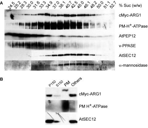
Figure 6.
Immunoblot Analyses of Protein Extracts from Transgenic arg1-2 Carrying the cMyc-ARG1 Construct, Showing the Association of ARG1 with Multiple Membranes.
(A) Fractionation of cMyc-ARG1, PM H+-ATPase (a plasma membrane marker), AtPEP12 (an endosomal marker of the vacuolar pathway), anti-vacuolar pyrophosphatase (v-PPASE; a vacuolar marker), AtSEC12 (an ER marker), and α-mannosidase (an early Golgi marker) in a 14 to 50% (w/w) sucrose gradient. The sucrose concentration of each fraction, as determined by its refractive index, is given at top. A total of 20 μL of each fraction was loaded per lane. The results shown are representative of two separate gradients.
(B) Fractionation of cMyc-ARG1, PM H+-ATPase, and AtSEC12 in the two-phase partitioning experiment. cMyc-ARG1 was found in both the plasma membrane–enriched upper phase (PM) and the plasma membrane–deprived lower phase (Others). The shift in mobility of cMyc-ARG1 in the plasma membrane fraction likely was an artifact of electrophoresis (see Results). A total of 75 μg of protein was loaded per lane. P150, microsomal membrane fraction; S150, soluble protein fraction.

