Figure 1.

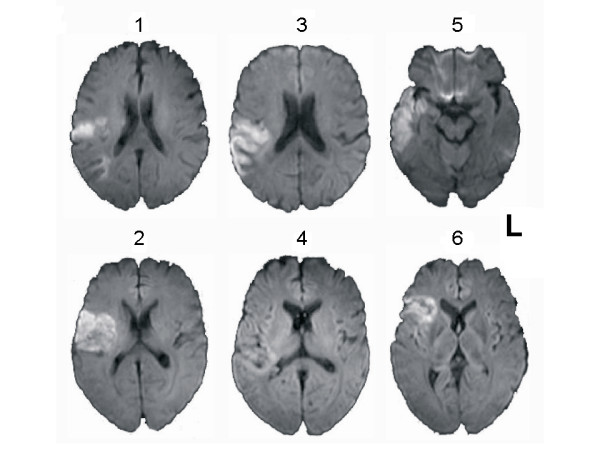
Ischemic Lesions. Axial Diffusion Weighted MR images of enrolled patients. The left side of the figure corresponds to the left side of the brain.

Ischemic Lesions. Axial Diffusion Weighted MR images of enrolled patients. The left side of the figure corresponds to the left side of the brain.