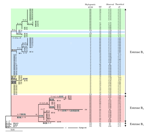
Figure 1.
Phylogenetic tree of Aes sequences from the 72 ECOR strains and 6 E. coli reference strains. The tree was reconstructed with PHYML [50]. E. fergusonii was used as an outgroup. Bootstraps are shown for values higher than 70%. Differences in amino acids are indicated on the branches. Differences for each branch were derived from comparison of consensus amino-acid sequences of the ancestors and descendants. Boxed amino-acid substitutions correspond to substitutions that change the overall pI of the protein. The phylogenetic groups A (blue box), B1 (green box), B2 (red box), D (yellow box) and ungrouped strains (UG) (white box), electrophoretic mobilities (Mf) obtained by polyacrylamide agarose gel electrophoresis [10] and the observed [10] and theoretical pI of Aes are indicated. nd: non determined. -: non significant results.

