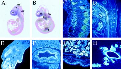
Figure 2.
LMO-4 expression pattern during mouse development by in situ hybridization. (A) Whole-mount in situ hybridization of e9.5, lateral view. (B) Whole-mount in situ hybridization of e10.5, lateral view. (C) Cross section at e10.5 displaying strong signal in dorsal root ganglia. (D) Sagittal section of the neck area at e14.5 showing strong expression in esophagus, vertebrae and thymus. (E) Section of a hindlimb showing expression in cartilage and developing epidermis. (F) Section from e17.5 showing expression in stomach. (G) Section from e17.5 showing expression in submandibular gland and in basal layer of the epidermis. (H) Section from a breast gland showing expression in epithelial cells in a 14.5-day pregnant mother. T, telencephalon; D, diencephalon; MS, mesencephalon; MT, metencephalon; N, nasal epithelium, OV, otic vesicle; BR, first brancial arch; F, forelimb.

