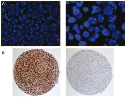
Figure 1.
(A) Fluorescence in situ hybridization on formalin-fixed, paraffin-embedded tissue sections on a tissue microarray. Left panel: interphase nuclei with BCL6 break apart (BCL6+). The most common signal pattern is one fusion signal (yellow) and two split signals (1 red, 1 green) visualized at 200× magnification. Right panel: BCL6+ interphase nuclei with break apart (split signals) and polyploidy (multiple fused signals) of the BCL6 locus (high power field). (B) Immunohistochemistry for BCL-6 on tissue microarray cores. Left panel: positive staining in virtually all large B cells. Right panel: negative staining.

