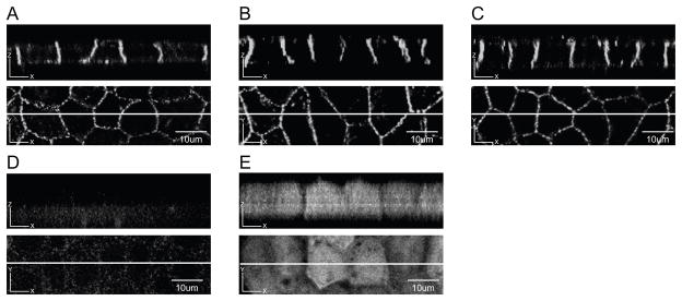
Fig. 3. WT Cx43-eYFP is selectively targeted to the basolateral domain of MDCK cells.
A,B,C. Three independent MDCK clones expressing WT Cx43-eYFP were cultured on filter inserts. Z sections of cell monolayers are shown in the top panels. Lines drawn through the XY plane in the bottom panels indicate the location of the Z sections. WT Cx43-eYFP was expressed on the basolateral membrane domain of MDCK cells. D. Z section of monolayer of uninfected MDCK cells showed background fluorescence. E. Z section of monolayer of MDCK cells expressing only eGFP showed a diffuse cytoplasmic pattern. Scale bar, 10 um.

