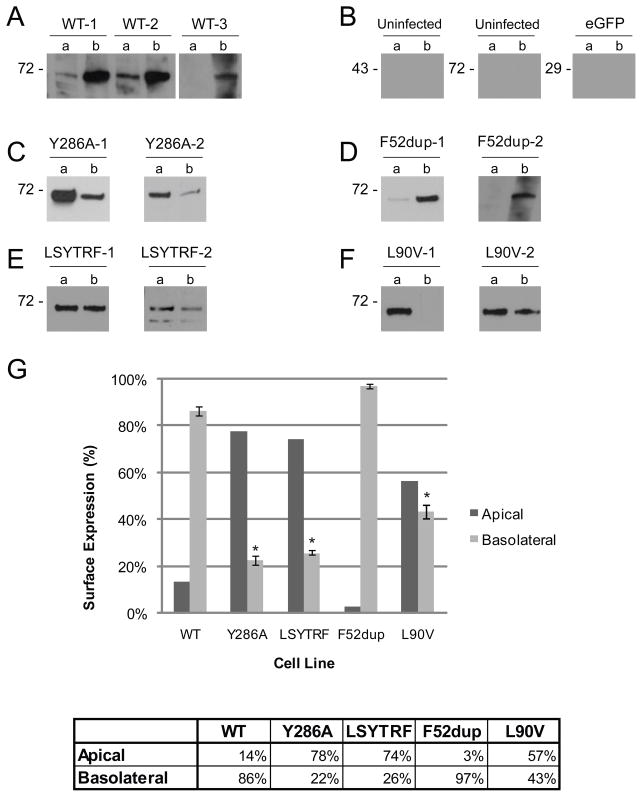
Fig. 6. Apical/basolateral cell surface distribution of Cx43-eYFP.
Selective surface biotinylation followed by Western blot analysis using the anti-Cx43 antibody showed that WT and the F52dup mutant construct were expressed predominantly on the basolateral surface (A, D), while the Y286A, LSYTRF, and L90V mutant constructs were not exclusively distributed on the basolateral surface (C, E, F). B. Uninfected MDCK cells showed no bands at ~70 kDa or 43 kDa. Absence of bands at ~29 kDa in control cells expressing only eGFP indicates that non-specific biotinylation of cytoplasmic protein did not occur. Two or three individual cell lines for each construct are shown. G. Surface expression as quantified by a fluorescence plate reader following selective surface biotinylation. The percent of signal found on the basolateral surface of Y286A, LSYTRF, and L90V was significantly different from that of WT (*). Error bars indicate s.e.m, n = 3–6 (varies for cell lines).

