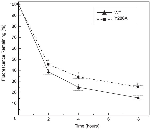
Fig. 8. The Y286A mutant construct shows impaired degradation as compared to WT.
Surface protein degradation assays on tissue culture dishes were conducted by labeling surface protein with membrane-impermeant sulfo-NHS-LC-biotin and lysing cells at 0, 2, 4, and 8 hours. eYFP fluorescence was quantified using a fluorescence plate reader. Fluorescence remaining (%) was calculated by normalizing to the reading at time 0 for each cell line. The Y286A mutation slightly impaired degradation of Cx43-eYFP from the surface as compared to WT. For each cell type, at least three independent experiments were performed on two clones. Data are graphed as mean ± s.e.m. (brackets represent s.e.m.).

