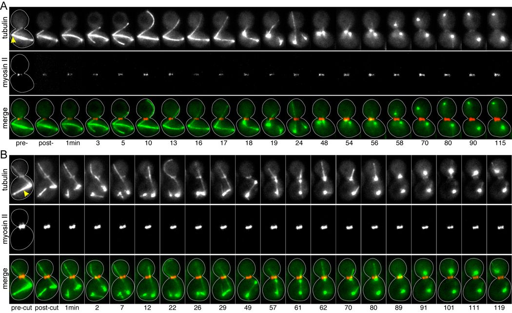
Figure 2.
Damaging the SPBs or spindle microtubules does not prompt mitotic exit. Timelapse images of microtubules labeled with GFP-tubulin and actomyosin rings labeled with RFP-myosin-II/Myo1, from movies of dyn1Δ mutant cells. Merge series depicts tubulin in green and myosin-II in red. Each image is a composite of 3 planes separated by 1µm. Strain: yJC6877. A) Ablation of the dSPB. Arrowhead marks the dSPB, which harbors the neck-interacting microtubule and was targeted for ablation. The spindle collapses within 18 min of irradiation. The actomyosin ring does not contract within 115 min, despite the migration of one of the resolved spindle poles into the bud at 56 min, indicating that this cell has not exited mitosis. B) Severing the spindle. The ablation beam was targeted to spindle microtubules, denoted by the arrowhead, resulting in the destruction of the spindle and resolution of the spindle poles. The spindle reforms by 29 min; however, this association is lost by 62 min. One spindle pole enters the bud at 89 min. The actomyosin ring does not contract within 119 min, indicating that this cell has not exited mitosis.

