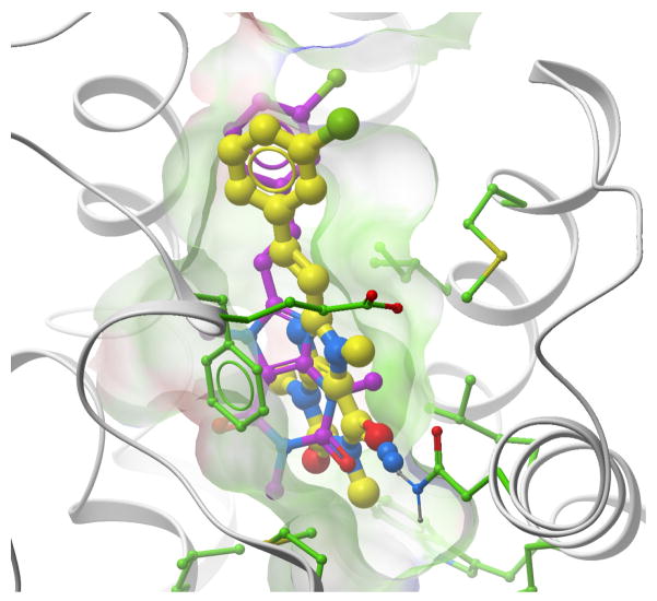
Figure 9.
A xanthine analogue (GLIDA ID#L011922) docked into the AA2AR. The crystal structure (3eml) of the receptor is shown by grey ribbon sticks with green carbons. The ligand conformation docked into the crystal structure has yellow colored carbon atoms, the conformation docked into the blindly predicted model (mod2upu) has magenta colored carbons.

