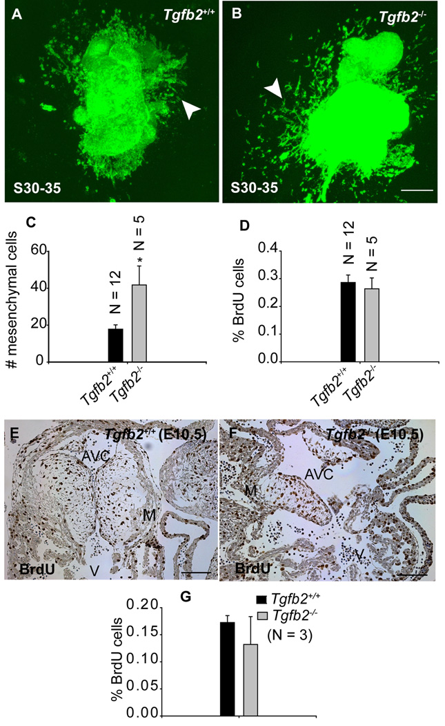
Fig.3. Effect of gene targeted disruption of Tgfb2 on cushion EMT and cell proliferation after the rudimentary cushions are formed at E10.5.
A-B: Representative confocal images of alexa-fluor488-phalloidin stained S30–35 (E10.5) AV canal explants of Tgfb2+/+ (A) and Tgfb2−/− (B) embryos after 60 hr of culturing. The AV explants in which the epicardial cell layer is removed or destroyed at the time of harvesting are cultured for 12 hr to let the pre-existing cushion mesenchymal cells migrate out of the AV explants and then transferred to fresh collagen gels for an additional 48 hr. (C) The number of mesenchymal cells (mean±s.e.m) below the gel surface (arrowhead, a-b). Notably, as the endocardium and epicardium are the only possible sources of mesenchyme in this experiment, differences between the wild type and Tgfb2−/− mice reflect the unimpeded endothelial source and, perhaps, a few cells that may have escaped the epicardial disruption procedure (see Experimental Procedures for details). (D) % of BrdU-incorporated mesenchymal cells (mean±s.e.m) in vitro on collagen gels. The number of mesenchymal cells is significantly increased in Tgfb2−/− mice (C) (*P=0.0032) but there is no difference in % of BrdU cells between wild-type and mutants (D). e-g: Anti-BrdU-stained sections of E10.5 AV cushions of wild-type (E) and Tgfb2−/− (F) mice and the % of BrdU-incorporating cushion cells (G) (brown color staining in E-F). Cell proliferation is comparable between wild-type and mutant cushions at E10.5 (P=0.9416). Images (E,F) are representative of three wild-type/mutant embryo pairs. Scale bar: 100 µm (A,B,E,F).

