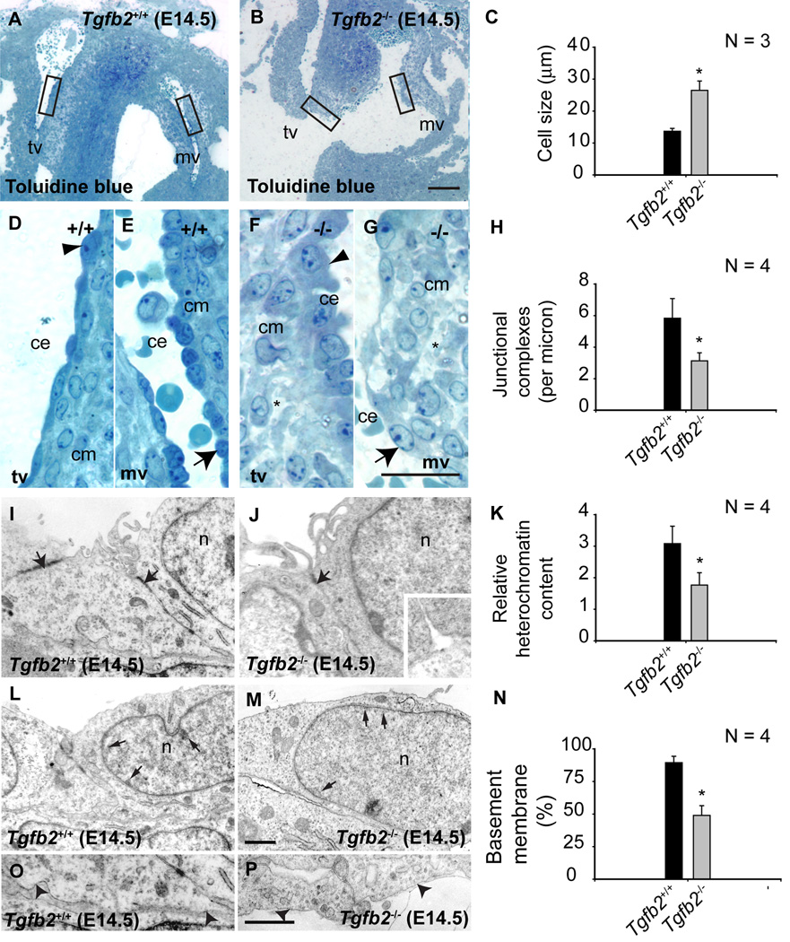
Fig.4. Cellular and ultrastructural changes in cushion endothelial cells in Tgfb2 −/− mice at E14.5.
A-G: Toluidine blue staining of wild-type (A) and Tgfb2−/− (B) AV cushions. Boxes in (A) and (B) are magnified in (D,E) and (F,G), respectively. Asterisks (F,G) indicate the abnormal organization and composition of cardiac jelly in mutants. Cushion endothelial cells are enlarged in mutants (arrowheads (D,F) and arrows (E,G)). C: Morphometric comparison showing significantly increased cushion endothelial cell size (mean±s.e.m) in Tgfb2−/− embryos (*P= 0.0138). I-Q: Ultrastructural and morphometric comparison of wild-type and Tgfb2−/− mice by transmission electron microscopy. Electron micrographs of representative E14.5 Tgfb2+/+ (I,L,P) and Tgfb2−/− (J,M,Q) mice, illustrating decreased endothelial junctional complexes (arrows, I,J), heterochromatin organization (arrows, L,M), and basement membrane (arrowheads, P,Q). Morphometric comparison (mean±s.e.m) of junctional complexes (H), relative heterochromatin content (K) and % of basement membrane (O) confirms these findings. *P=0.0320 (junctional complexes), 0.0306 (heterochromatin content), 8.8927e-5 (basement membrane). Images (A,B,D-G) and (I,J,L,M,P,Q) are representative of four wild-type/mutant embryo pairs. Scale: 100 µm (a,b), 25 µm (D,E,F,G), 1 µm (scale in Q applies to I,J,L,M,P). mv, mitral valves; tv, tricuspid valves; ce, cushion endothelium; cm, cushion mesenchyme; n, nucleus.

