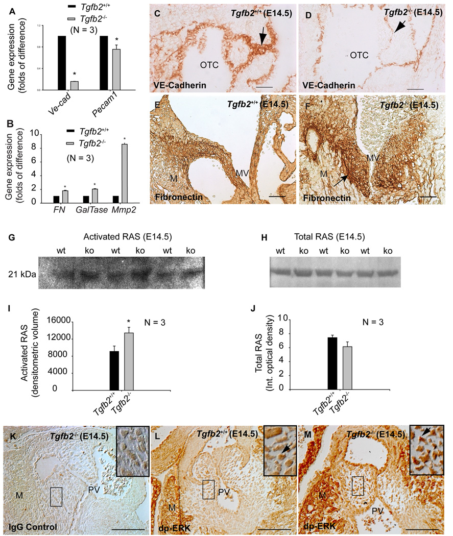
Fig.5. Cushion EMT marker expression in Tgfb2−/− mice at E14.5.
A-B: Q-RT-PCR (mean±s.e.m.) showing significantly decreased VE-Cad (*P=7.0000e-10) and Pecam1 (*P=0.0392) (A), and increased Fibronectin (*P = 0.0016), GalTase (*P=2.6790e-4) and Mmp2 (*P=6.4998e-6) (B) in AV explant tissue of Tgfb2−/− mice. Each wild-type value is normalized to 1.0. C-D: Anti-VE-Cadherin staining of wild-type (C) and Tgfb2−/− (D) mice shows that VE-Cadherin is reduced in mutants (arrows, C-D). E-F: Anti-FN staining of wild-type (E) and Tgfb2−/− (F) mice confirms the increased FN in AV cushions of Tgfb2−/− mice (arrow, F). Note that Tgfb2−/− cushions in (F) are naturally enlarged. G,H: Representative western blots showing increased activated RAS (G) and unaltered total RAS (H) in Tgfb2−/− hearts. The blots are a representative of at least 3 wild-type/mutant embryo pairs. I,J: Densitometric quantification (mean±s.e.m) confirms increased activated RAS in mutants (I) (*P=0.0394) without any significant change in the total RAS (J) (P=0.1710). K-M: Tgfb2−/− cushions stained with IgG (K) and anti-dp-ERK (L) are compared with anti-dp-ERK stained cushions of Tgfb2+/+ mice (M). Areas under the box in (K–M) are magnified (insets). Tgfb2−/− cushions have increased dp-ERK (arrows, L-M). Images (C-D, E-F, K-M) are representative of at least three wild-type/mutant embryo pairs. Scale bar: 50 µm (C-D, E-F, K-M). OTC, outflow tract cushion; MV, mitral valves; M, myocardium; PV, pulmonary valves.

