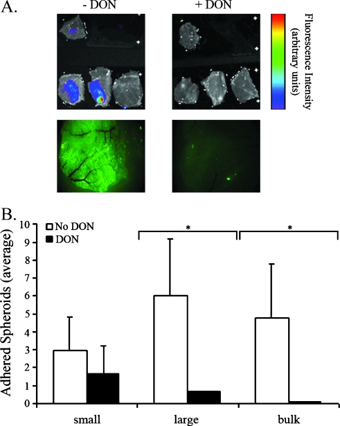
Figure 4.
Peritoneal adhesion is mediated by HA. CD-1 nude OVX mice were treated with DON (i.p.; 7.5 µg/g body weight every 24 hours for 2 days). DON was added also to the spheroid culture medium (5 µg/ml every 24 hours for 2 days). Control mice and spheroids were not treated with DON. DON-treated and control spheroids (0.8–1 mm in diameter) were stained with 4-Di-10-Asp and i.p. inserted into DON-treated and control CD-1 nude OVX mice. Adhesionwas viewed and quantified 9.5 hours after insertion. (A) Top, color-coded fluorescent images of equally excised peritonea (overlaid on a grayscale photograph). The single upper peritoneum in each image represents fluorescence background from a naive mouse. Bottom, fluorescent images (Zeiss) of whole peritonea. (B) Average number of adhered spheroids in equally excised peritonea (DON-treated, black, n = 5; control, white, n = 3). Small, ≤0.1 mm; large, >0.1 mm; bulk, >1 spheroid (error bars, SE; *P < .05, two-tailed, unpaired t test).

