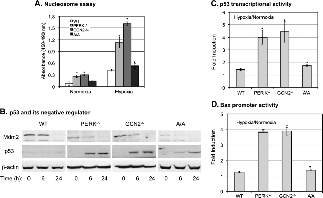
Figure 3.
Both PERK and GCN2 regulate p53 signaling cascade and apoptosis in hypoxia. MEFWT, MEFPERK-/-, MEFGCN2-/-, and MEFA/A cells were used in the experiments. (A) The cells were exposed to normoxia or hypoxia for 36 hours before apoptotic assays by detecting cleaved histone/DNA complex. The bars represent the means of three independent experiments. *P < .05 mutant versus wild type under normoxia or hypoxia. (B) The cells were treated with hypoxia for the time points as indicated and the protein level of Mdm2 was analyzed by Western blot. (C and D) The cells were cotransfected with p53 luciferase reporter plasmid or Bax luciferase reporter plasmid. Renilla luciferase reporter plasmid was used to normalize the transfection efficiency. Twenty-four hours after transfection, the cells were exposed to normoxia or hypoxia for 24 hours, and the activities of p53 and Bax were analyzed by luciferase assay. The bars represent the means of three independent experiments. *P < .05 mutant versus wild type.

