Figure 4.
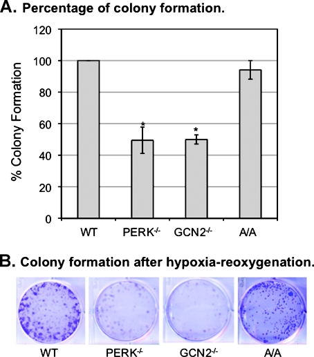
Both PERK and GCN2 mediate recovery of cells from hypoxic stress. MEFWT, MEFPERK-/-, MEFGCN2-/-, and MEFA/A cells were exposed to hypoxia for 24 hours. Cells were collected and stained by Trypan blue. Nonapoptotic cells were counted. Five thousand cells were replated and cultured under normoxia for 6 days. Cells were then fixed by methanol for 10 minutes at -20°C and stained by 1% crystal violet (25% methanol) for 10 minutes, washed by distilled water, and dried. (A) The colonies with a size greater than 0.5 mm were counted. The degree of colony formation was expressed as percentage of MEFWT cells. The bars represent the means of three independent experiments. *P < .05 mutant versus wild type. (B) The plates were photographed using microscopy equipped with a Nikon digital camera.

