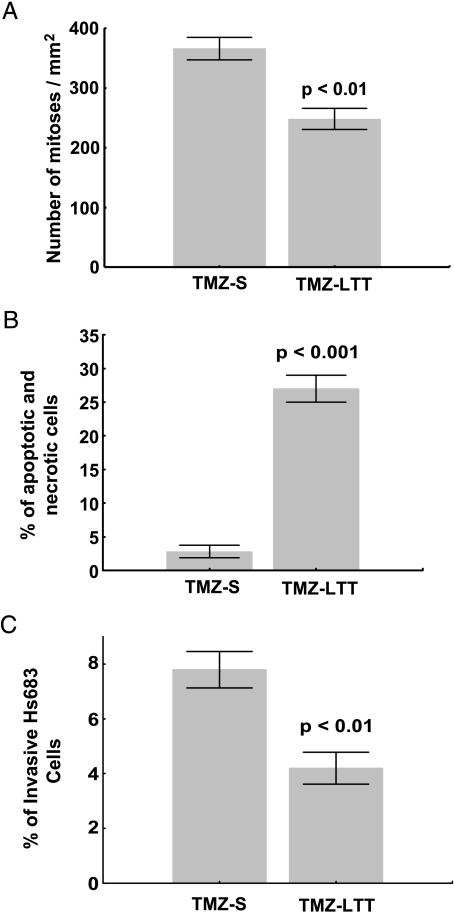
Figure 5.
(A) Quantitative determination of the percentage of mitotic cells during 72 hours in Hs683 oligodendroglioma cell populations that have been left untreated (TMZ-S Hs683 cells) or that have been able to grow in vitro over months in the presence of 1 mM TMZ (TMZ-LTT Hs683 cells; see Materials and Methods). (B) Flow cytometry analysis of double-stained (Annexin V and PI) TMZ-S and TMZ-LTT Hs683 cells. Apoptotic cells include annexin V+/PI- (early apoptosis) and annexin V+/PI+ (late apoptosis), whereas necrotic cells are annexin V-/PI+ and normal cells annexin V-/PI-. (C) Characterization of the invasiveness of TMZ-S and TMZ-LTT Hs683 cells cultured for 24 hours in Matrigel-coated Boyden chambers. Data are illustrated in A to C as mean ± SEM values, and all experiments have been carried out in triplicate.

