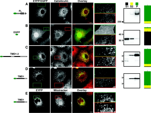
Figure 2. N-terminally truncated IP3R1 fragments are targeted to the ER by the first TMD pair.
COS-7 cells transiently transfected with the indicated constructs are shown in the left column and stained for calreticulin [MitoTracker in (E)] in the second column. The third column shows the first two columns overlaid with the construct in green and calreticulin (or MitoTracker) in red. Bars, 10 μm. The fourth column shows enlargements of the highlighted boundaries, with green and red borders enclosing the construct and organelle marker respectively. Here, and in all subsequent Figures, images are representative of at least three independent transfections. The fifth column shows Western blots (with an antibody to GFP) of the three fractions derived from subcellular fractionation of the cells: S1 (first supernatant; cytosolic proteins), S2 (second supernatant; peripheral membrane proteins) and P (pellet; integral membrane proteins). For each gel, the three lanes were loaded with material from an equivalent number of cells. Molecular-mass markers are shown in kDa. The final column summarizes the results obtained from the subcellular fractionation (values are means±S.E.M., n≥3; see the Experimental section).

