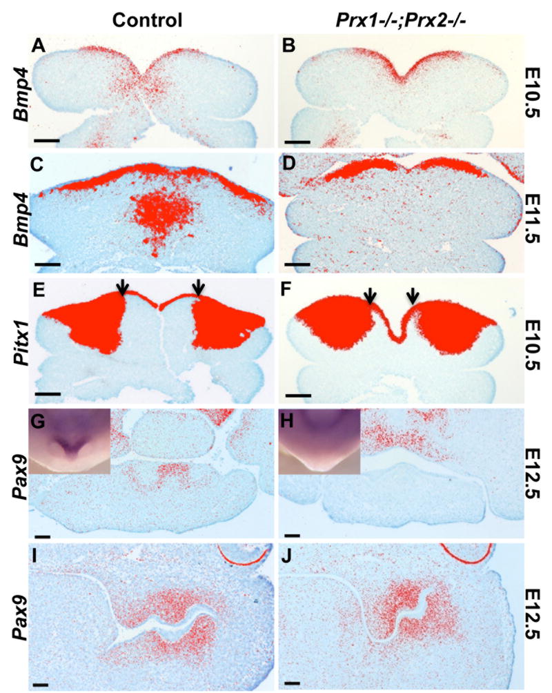
Figure 3. Effects of absence of Prx on the expression of Bmp4, Pitx1 and Pax9.
A-D are pseudo colored images of in situ hybridization analysis with Bmp4 on transverse sections of mandibular processes of control (A, C) and Prx1-/-;Prx2-/- (B, D) embryos at E10.5 (A, B) and E11.5 (C, D). The expression of the Bmp4 in the mesenchyme of the medial region is reduced in the Prx1/Prx2 double mutants as compared to control. There are no significant changes in the expression of Bmp4 in the mandibular epithelium between the two genotypes.
E, F are pseudo colored images of in situ hybridization analysis with Pitx1 on transverse sections of mandibular processes of control (E) and Prx1-/-;Prx2-/- (F) embryos at E10.5. The expression of Pitx1 in the mandibular epithelium is similar between the control and Prx1/Prx2 double mutants. The expression of the Pitx1 in the mandibular mesenchyme in extended into the medial region in the Prx1/Prx2 double mutants as compared to control.
G-J are pseudo colored images of in situ hybridization analysis of Pax9 expression in the dental papillae of the mandibular incisors (G, H) and molars (I, J) in transverse sections of control (G, I) and Prx1/Prx2 double mutant (H, J) mandibles at E12.5. Note Pax9 expressed in the dental papillae of the mandibular incisors in the control (G), but not in the Prx1/Prx2 double mutants (H). Whole-mount in situ hybridization analysis with Pax9 is shown in the insets. Note that Pax9 is expressed in the rostral region of the control mandible but not in Prx1;Prx2 double mutants. Also note Pax9 is expressed in similar domain and intensity in the dental papillae of the mandibular molars in the control (I) and the Prx1/Prx2 double mutants (J). Scale bars =100 um.

