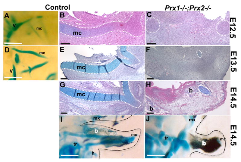
Figure 5. Effect of absence of Prx genes on development of Meckel’s Cartilage.
Whole mount staining (A, D, I, J) and histological analysis (B, C, E, F, G, H) of longitudinal sections of mandibular processes from control (A, B, D, E, G, I) and Prx1;Prx2 double mutant (C, F, H, J) embryos at E12.5 (A-C), E13.5 (D-F) and E14.5 (G-J). In all images the symphyseal portion of the Meckel’s cartilage and the medial region of the developing mandible is on the right.
Side view (A) and longitudinal section (B) of the mandibular processes from control embryos, showing the presence of two rods of Meckel’s cartilage at E12.5. The dashed white line oulines the forming Meckel’s cartilage. C is a longitudinal section of a mandibular process from Prx1;Prx2 double mutant embryo at E12.5 showing the absence of rods of Meckel’s cartilage. The dashed white circles outlines the areas of condensation for the most rostral and caudal ends of Meckel’s cartilge.
Side view (D) and longitudinal section (E) of the mandibular processes from control embryos showing the presence of the two rods of Meckel’s cartilage (outlined by dashed white line) at E13.5. F is a longitudinal section of a mandibular process from Prx1;Prx2 double mutant embryo at E13.5 showing the absence of rods of Meckel’s cartilage. Note the small remnants of Meckel’s cartilage (indicated by dashed white circle) in the rostral region of the mandibular processes of Prx1;Prx2 double mutant at E13.5.
(G) Longitudinal section through the mandibular process isolated from E14.5 control embryo showing the fully formed rod of Meckel’s cartilage (outlined by dashed white line). (H) Longitudinal section through the mandibular process from Prx1;Prx2 double mutant embryo at E14.5 showing the absence of the fully formed rod of Meckel’s cartilage. Note the remnant of Meckel’s cartilage (indicated by dashed white circle) in the rostral region of the mutant mandibular process.
I and J are side views of stained head from control (I) and Prx1;Prx2 double mutant (J) embryos at E14.5 showing the formation of mandibular bones stained with Alizarin Red (indicated by b). Note the increased bones stained with Alizarin Red in the mandibular processes of Prx1;Prx2 double mutant. The soft tissue of the face is highlighted by dashed black lines. Abbreviations: b, mandibular bones; h, hyoid; mc, Meckel’s cartilage; mx, maxilla; tr, tympanic ring; v, vertebrate column.Scale bars =100 um.

