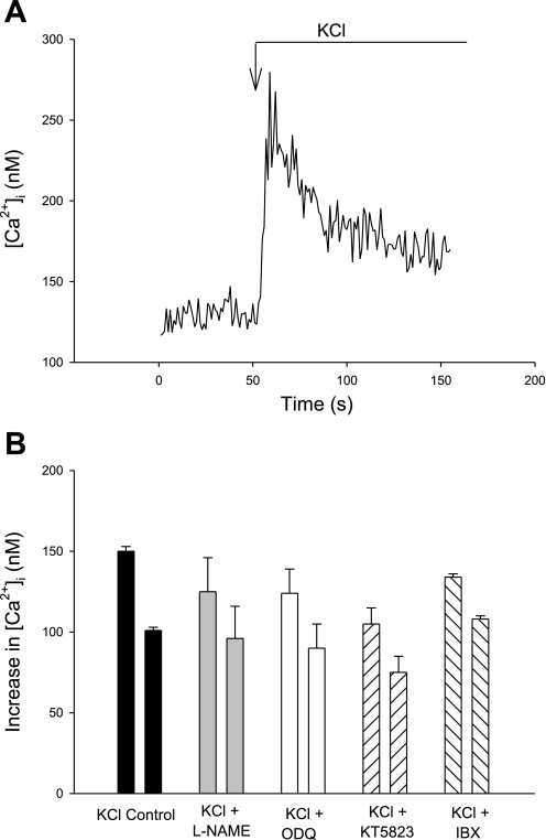
Fig. 1.
Changes in cytosolic Ca2+ concentration ([Ca2+]i) of isolated afferent arterioles to KCl (50 mM). A: representative recording of [Ca2+]i response to KCl. B: summary data of increases in peak (left bar in each pair) and plateau (right bar in each pair) [Ca2+]i in afferent arterioles in response to KCl (50 mM) in afferent arteriolar vascular smooth muscle cells (VSMC) in the absence or presence of the inhibitors Nω-nitro-l-arginine methyl ester (l-NAME; nitric oxide synthase), 1,2,4-oxodiazolo-[4,3-a]quinoxalin-1-one (ODQ; soluble guanylyl cyclase), KT-5823 (protein kinase G), or iberiotoxin [IBX; Ca2+-dependent big potassium channel (BKCa2+)]. P = not significant (NS) vs. control for each agent, both peak and plateau. See Table 1 for P and n values.

