Fig. 1.
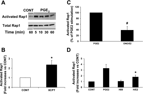
PGE2 activates Rap via EP4 and PKA. Neonatal ventricular myocytes (NVMs) were treated with or without 1 μM PGE2 for 0–60 min and then lysed for the Rap1 activation assay as described in methods. A: time course of Rap1 activation. Representative Western blots of activated Rap compared with total Rap protein are shown. Data represent 2 separate experiments. B: Rap activation via Epac. NVMs were treated for 5 min with 60 μM 8CPT-2Me-cAMP (8CPT). Activated Rap is expressed as the fold increase compared with the vehicle-treated control (CONT). *P ≤ 0.05 vs. the control. C: EP4 dependence of Rap activation. NVMs were treated with PGE2 for 5 min after a 1-h pretreatment with or without 10 μM ONO-AE3-208 (ONO). The graph is a summary of data, which are expressed as the percentage of PGE2 stimulation. Bars are means ± SE from 5 separate experiments. #P ≤ 0.01 vs. PGE2. D: PKA dependence of Rap activation. NVMs were treated for 5 min with PGE2 after a 1-h pretreatment with 5 μM H89 (H). Activated Rap is expressed as the fold increase compared with the control, which was treated with DMSO and ethanol vehicle. Data are means ± SE from 5–9 separate experiments. *P ≤ 0.01 vs. PGE2.

