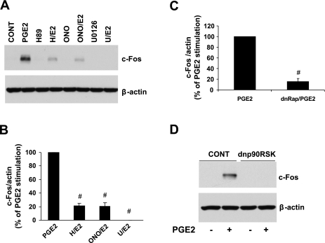
Fig. 5.
EP4, PKA, Rap, ERK1/2, and p90RSK regulate c-Fos expression. A: representative Western blots showing the effect of ONO, H89, or U-0126 on the PGE2 stimulation of c-Fos. Cells were pretreated for 1 h, treated with PGE2 for 60 min, and then lysed for the detection of c-Fos by Western blot analysis. B: summary graph of data from 4 to 6 separate experiments. c-Fos normalized to β-actin is expressed as a percentage of PGE2 stimulation. #P ≤ 0.01 vs. PGE2. C: Rap involvement. NVMs were transfected with dnRap for 24 h, treated with PGE2 for 60 min, and then lysed for Western blot analysis. The graph is a summary of data from 3 separate experiments, and the data are expressed as a percentage of PGE2 stimulation. #P < 0.01 vs. PGE2. D: involvement of p90RSK in the PGE2 regulation of c-Fos. NVMs were transduced with dnp90RSK adenovirus or LacZ control virus for 48 h, treated with PGE2 for 60 min, and then lysed for Western blot analysis. Blots are representative of 3 separate experiments.

