Fig. 7.
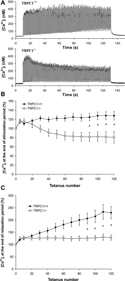
Involvement of TRPC1 in muscle fatigue: [Ca2+]i transients. A: examples of [Ca2+]i transients measured in flexor digitorum brevis (FDB) fibers from TRPC1+/+ (top) and TRPC1−/− mice (bottom) submitted to the fatigue protocol described in Fig. 6. B: quantification of maximal tetanic [Ca2+]i during the protocol of fatigue ([Ca2+]i measured at 1st, 5th, and then every 10th tetanus). Results are expressed relative to maximal [Ca2+]i obtained during 1st tetanus. C: quantification of the [Ca2+]i obtained at the end of each relaxation period during the protocol of fatigue. ([Ca2+]i measured at 1st, 5th, and then every 10th tetanus). Results are expressed relative to maximal [Ca2+]i obtained during 1st tetanus. Statistical analysis: *TRPC1+/+ (n = 13) different from TRPC1−/− (n = 22), P < 0.05, 1-way repeated-measures ANOVA followed by Bonferroni multiple-comparison test.

