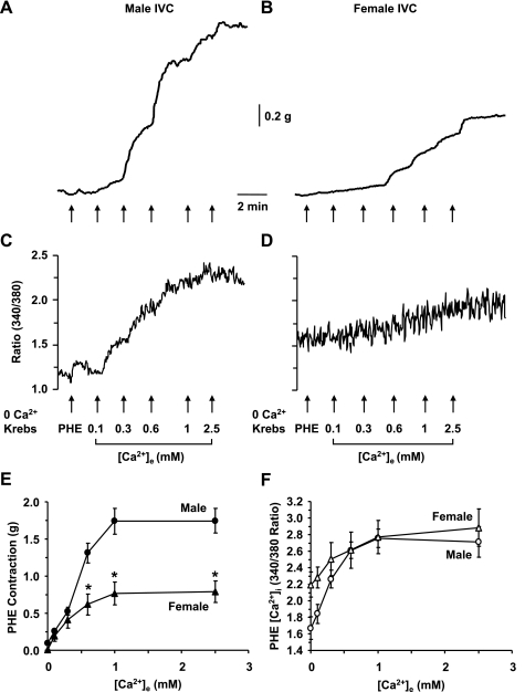
Fig. 3.
PHE-induced [Ca2+]e-contraction and [Ca2+]e-[Ca2+]i curves in male and female rat IVC. IVC segments from male (A, C) and female rats (B, D) were incubated in Ca2+-free (2 mM EGTA) Krebs for 5 min, then nominally 0 Ca2+ Krebs for 5 min. The tissues were stimulated with PHE (10−5 M), and the initial contraction (A, B) and [Ca2+]i (C, D) were recorded. Increasing concentrations of extracellular CaCl2 (0.1, 0.3, 0.6, 1, 2.5 mM) were added, and the PHE-induced [Ca2+]e-contraction curve (E) and [Ca2+]e-[Ca2+]i curve (F) were constructed and compared in male and female IVC. Data are expressed as means ± SE; n = 4. *Measurements in female IVC are significantly different compared with corresponding measurements in male IVC (P < 0.05).

