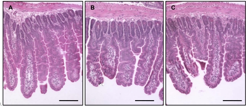
Fig. 2.
Light micrograph of a cross section (hematoxylin and eosin stained) of proximal jejunum of mice fed with control diet and killed at day 8 postirradiation: 0 Gy (A), 8.5 Gy (B), and 10 Gy (C). Bars are 75 μm. Villi were intact in unirradiated and irradiated mice, independent of diet. Mice irradiated with 7 Gy and mice consuming vitamin A-supplemented diet not shown.

