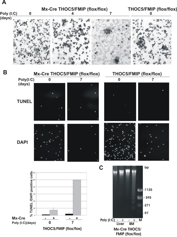
Figure 6.
Deletion of THOC5/FMIP causes apoptosis of leukocytes in bone marrow. Six-week-old Mx-cre THOC5/FMIP (flox/flox) mice (n = 9) and THOC5/FMIP (flox/flox) control mice (n = 6) were injected with poly (I:C) (250 μg each) and the femora were then isolated. (A): Bone marrow cells were spun down onto glass slides and then stained with May-Grunwald Giemsa and hematoxylin. (B): Aliquots of same preparation were stained with TUNEL and DAPI. Results are the mean +/- SEM of %TUNEL positive/DAPI positive cells (n >2000 cells). Original magnification: ×200 for all panels. (C): Aliquots of 2-3 μg of DNA from liver and bone marrow of poly (I:C) treated (+) and untreated (-) Mx-cre THOC5/FMIP (flox/flox) mice were separated on 1.5% (w/v) agarose gel, stained with ethidium bromide (2 μg/ml) and photographed under UV light. M: base pair Marker.

