
Figure 3.
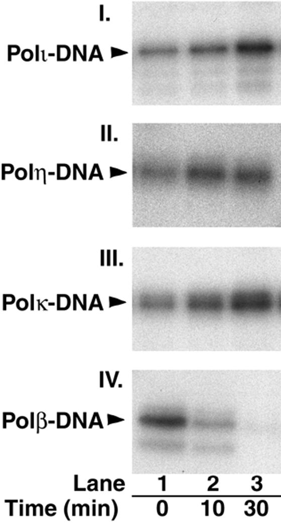
Time course of polymerase–DNA cross-linking by borohydride. Polι (panel I), Polη (panel II), Polκ (panel III), or Polβ (panel IV; each at 10 nM) were mixed at 0°C with the internal 5′-dRP-containing 3′-32P-labeled DNA (50 nM) shown in Figure 2A, followed by incubation at 37°C for 0, 10, or 30 min (lanes 1–3, respectively) before the addition of 20 mM NaBH4 and further incubation at 0°C for 30 min. After the addition of SDS-containing loading buffer, the samples were resolved on an 8% SDS–polyacrylamide gel, and the cross-linked polymerase–DNA products, indicated by arrows on the left of panels, were analyzed by autoradiography.