Figure 3.
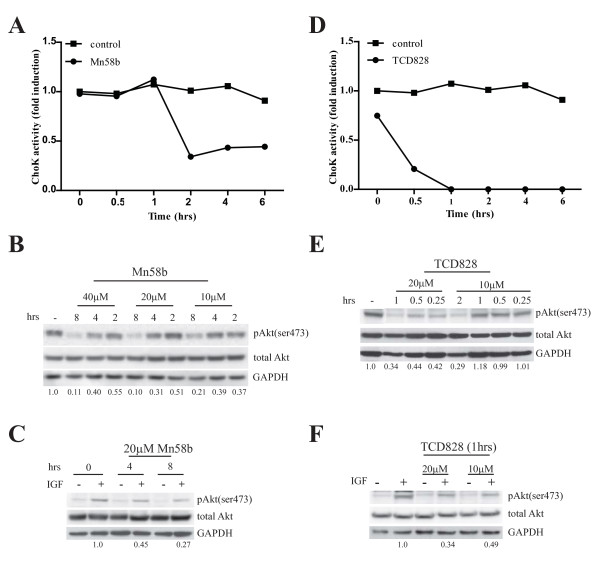
ChoK inhibitors inhibit ChoK activity and Akt phosphorylation. A, MDA-MB 468 cells were treated with 20 μM Mn58b for indicated time. Lysates were harvested and ChoK activity determined as described in methods. B, MDA-MB 468 and C, serum-starved MDA-MB 231 were treated with Mn58b for indicated time with the specified concentration. MDA-MD 231 cells were stimulated with IGF for 15 mins. 30 μg cell lysate were subjected to western blot with the indicated antibodies. D, MDA-MB 468 cells were treated with 20 μM TCD828. ChoK activity was determined as described in methods. E, MDA-MB 468 and F, serum starved MDA-MB 231 were treated with TCD828 for the indicated time with the specified concentration. MDA-MD 231 cells were stimulated with IGF for 15 mins. 30 μg cell lysate were subjected to western blot with the indicated antibodies. pAkt(ser473) signals were quantified using Image J program and normalized to respective total Akt signal. Values below the blots indicate the normalized Akt(ser473) phosphorylation compared to untreated control.

