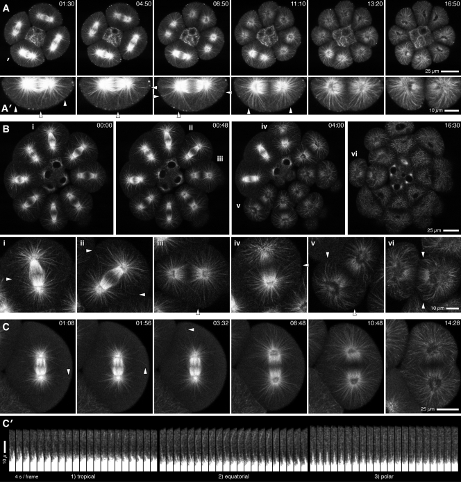
Figure 1.
Microtubules in live urchin embryos. All panels show single confocal sections. (A and A′) 16-cell purple urchin embryo; A′ shows a 2× enlarged view of the lower cell (indicated by white mark in A). Microtubules approach the cortex everywhere before anaphase onset (1 min, 30 s); during anaphase, just before furrowing, many astral microtubules penetrate both polar and equatorial cortex (arrowheads in A′). (B) Vegetal view, 28-cell sand dollar embryo; i–vi are 2× enlarged views as indicated. Astral microtubules frequently cross spindle midplane before and during anaphase (i–iii), approach within 1 µm of the equatorial surface before furrowing (ii–iv), and curve inward as the furrow ingresses (v and vi). Arrowheads point to exemplars. (C) Eight-cell sand dollar embryo; single microtubules grow as far as the cell surface in all directions (equatorially in the 01:56 frame, tropically in the 01:08 frame, and toward the pole in the 03:32 frame). Astral microtubules reach the polar cortex most densely in anaphase (08:48) but also reach the equator before furrowing (frame 10:48). (C′) Enlargement of successive frames for microtubules indicated by arrowheads in C (intensities squared to enhance contrast). Video 1 corresponds to A–C. Time is indicated in minutes:seconds.

