Figure 6.
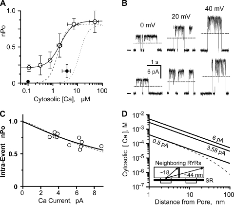
Flux inhibition of coordinated RYR2 channel activity. (A) Cytosolic Ca2+ sensitivity of coordinated RYR2 channel activity (nPo). Overall nPo was defined from coordinated channel recordings at 0 mV. The cytosolic solution contained 1 mM EGTA and Tris-HEPES (120/250 mM; pH 7.4). The luminal solution contained Ca-HEPES (50/250 mM; pH 7.4). Open circles summarize the nPo (mean ± SEM; n = 7 different experiments). The solid line is a Hill equation fit to open circles (EC50 = 1.98 µM, PoMax = 0.86, PoMin = 0.26, and Hc = 1.7). Small filled circles are after cytosolic Mg-ATP was added (1 mM of free Mg2+ and 5 mM of total ATP; n = 5 different experiments). Thin dashed and dotted lines are the single RYR2 50L and 50L plus Mg-ATP curves from Fig. 1 B. (B) Sample recording of coordinated events (two channels present). Solutions here are like those described for A (0.1 µM of cytosolic free Ca2+). Sample events at 0, 20, and 40 mV are shown. Open events are upward, zero current level is marked at left, and one open-channel current level is indicated by the dashed lines. All recordings shown are from the same channel incorporation. (C) Inhibition of intra-event nPo by increasing Ca2+ flux. Results collected from five different coordinated channels where only two channels were present. Solutions like those described for A (0.1 or 0.3 µM of cytosolic free Ca2+). Unit current was varied by changing membrane potential. Open circles are individual determinations. Dashed line is reproduced from Fig. 3 C. Thick solid line is a fit to the open circles and indicates a site distance from the pore of 1.5 nm (IC50 = 6.2 mM and Hc = −1.3). The free bath Ca2+ used for this fitting was 134 µM, which is the average predicted free [Ca2+] (for 3.58 and 6.20 pA) at 30 nm from an open pore. This distance (30 nm) is approximately the inter-pore distance of two adjacent RYR2 pores in cells. (D) Calcium diffusion from a point source was calculated with cytosolic Ca2+ of 100 nM and no buffer present (thick lines) for unit Ca2+ currents of 6, 3.58, and 0.5 pA. The dashed line is for 0.5 pA current with 244 µM of cytosolic Ca2+ buffer (Km = 673 nM) present (Bers, 2001). It was assumed that the kON of this buffer was diffusion limited. (Inset) A cartoon depicting salient dimensions of neighboring RYR2 channels.

