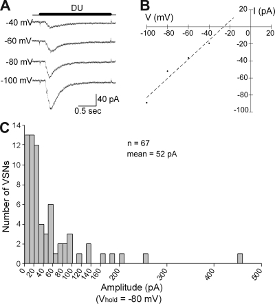
Figure 1.
Isolated mouse VSNs responded to dilute urine. (A) Perforated patch clamp recordings from the soma showed inward currents induced by a 2-s application of urine at voltage steps from −100 to −40 mV (20-mV increments). The time for the urine application is shown by the dark line above current traces. DU, 1:500 dilute urine. (B) I-V plot for the urine response of the VSN shown in A. Dashed line shows the best-fit line (y = 1.1183x plus 29.118; R2 = 0.9521). (C) Summary of the peak amplitudes for urine-induced inward currents at Vhold = −80 mV for 67 urine-responsive VSNs (one to two cells per mouse).

