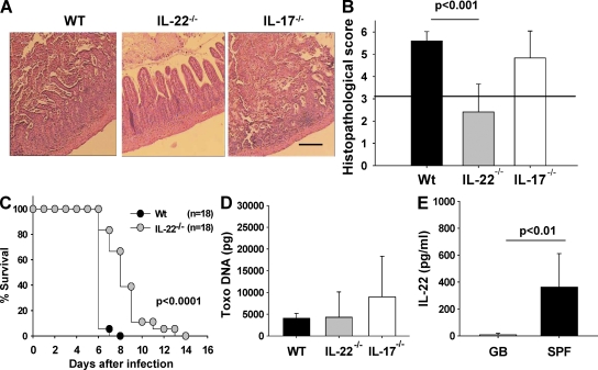
Figure 5.
IL-22–deficient mice are resistant to T. gondii–induced immunopathology. (A) Histopathology of hematoxylin and eosin–stained ileal sections of WT, IL-22−/−, and IL-17−/− mice 8 d after infection. (B) Histopathology scores of the ileum of WT, IL-22−/−, and IL-17−/− mice 8 d after infection. The horizontal line indicates the border between mild inflammation (<3) and necrosis (>3). (C) Survival of WT and IL-22−/− mice after oral infection with 100 cysts of T. gondii. (D) T. gondii DNA concentration in ileum of WT, IL-22−/−, and IL-17−/− mice 8 d after infection. (E) IL-22 concentration in culture supernatants of ileal biopsies from gnotobiotic (GB) and specific pathogen-free (SPF) mice 8 d after infection (ELISA). Data (one representative out of three independent experiments) from three to five mice per group are given as means ± SD, and p-values were determined by the Mann-Whitney U test. Bar, 100 µm.

