Figure 2.
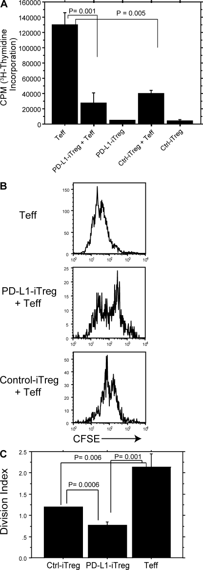
PD-L1–induced CD4+ Foxp3+ T reg cells suppress CD4+ T eff cells in vitro. (A) PD-L1 iT reg cell function was assessed by [3H]thymidine incorporation of naive CD4+CD25− T eff cells after 3 d of co-culture at a 1:1 T reg/T eff cell ratio plus PD-L1 beads (5:1 bead/T eff cell ratio). Data represent the mean ± SD and are representative of at least two independent experiments. (B) PD-L1 iT reg cell function was assessed by CFSE dilution of naive CD4+CD25− T eff cells after 3 d of incubation with 1:1 T reg/T eff cell ratio and PD-L1 beads (5:1 bead/T eff cell ratio). Data represent the mean ± SD and are representative of three similar experiments. (C) Quantification of T eff cell proliferation in B, analyzing the division index of gated CD4+CD45.1+ (the number of divisions a single cell has divided) by FlowJo software. Data represent the mean ± SD.

