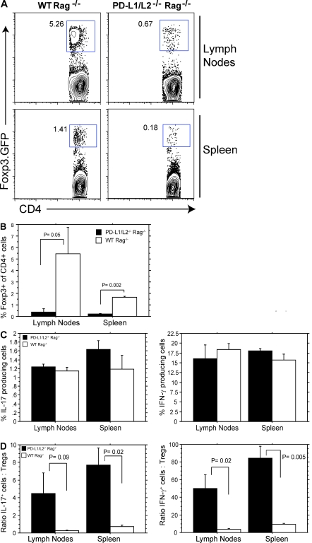
Figure 5.
Attenuated iT reg cell development in PD-L1−/−PD-L2−/− mice in vivo. (A) CD4+CD62LhiFoxp3.GFP− cells were adoptively transferred i.v. into the tail veins of WT Rag−/− or PD-L1−/−PD-L2−/−Rag−/− mice. Spleens and lymph nodes were analyzed for Foxp3.GFP expression 14–17 d after transfer. (B) Quantitation of Foxp3.GFP expression from independent mice depicted in A. Data represent the mean ± SE of five independent mice. (C and D) Analysis of IL-17+ and IFN-γ+ T eff cells by intracellular cytokine staining (C) and ratios of IL-17–producing T eff/T reg cells and IFN-γ–producing T eff/T reg cells from WT Rag−/− or PD-L1−/−PD-L2−/−Rag−/− mice 14–17 d after transfer (D). Data represent the mean ± SD of n = 5 mice per group and are representative of two independent experiments.

