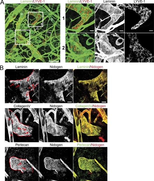
Figure 2.
Discontinuous expression of BM components on initial lymphatic vessels. (A) Confocal images of whole-mount ear skin preparations. BMs were identified by staining for laminin (green) and lymphatic vessels were identified by staining for LYVE-1 (red), respectively. The following tissue structures can be distinguished: a, arteriole; b, vein; c, capillary; d, fat cell; e, lymphatic vessel. Bar, 100 µm. Insets 1 and 2 show higher magnification pictures of respective areas on initial lymphatics. Bar, 20 µm. (B) Co-localization of major BM components on initial lymphatics. Lymphatic vessel edges are outlined red. Note the clear contours of BM perforations (arrows). Asterisks show areas on a lymphatic vessel (top row) and blood vessel (middle row), which do not represent perforations but rather tangential sections of the vessels caused by the limited z volume. Bar, 20 µm. Images are representatives of one of at least 11 different tissues investigated.

