Figure 3.
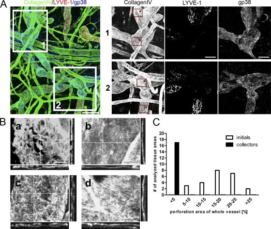
Collecting lymphatic vessels show continuous BM. (A) Confocal images of whole-mount ear skin preparations. BMs were identified by staining for collagen IV (green) and initial lymphatic vessels were identified by staining for LYVE-1 (red) and gp38 (blue). Collectors were identified by the lack of a LYVE-1 staining, but preserved gp38 staining. Bar, 100 µm. Insets 1 and 2 show higher magnification images of respective areas on collecting lymphatics. Bar, 50 µm. (B) Enlargements of insets in A demonstrating the discontinuous BM assembly on initial lymphatics in a and c in stark contrast to BM assembly on collector lymphatic vessels (b and d). Note the even distribution of collagen IV in b and d orthogonal plane views compared with a and c. Bar, 20 µm. (C) Quantification of perforation area in relation to total lymphatic vessel area for initial and collector lymphatics. Every single vessel area analyzed was grouped according to the fraction of the BM perforation area of the whole vessel area analyzed. Images are representatives of one of at least nine different tissues investigated. For the quantification in C, 24 areas in 11 different tissues were analyzed for initial lymphatic vessels and 17 areas in 9 different tissues were analyzed for collector lymphatic vessels.

