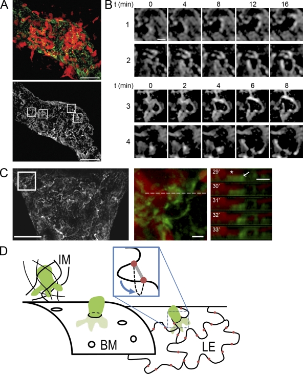
Figure 6.
DCs enter vessel lumen via endothelial flap valves. (A, top) DCs (red) entering an initial lymphatic vessel stained with LYVE-1 (green), which highlights the outline of lymphatic endothelial cells. (bottom) LYVE-1 staining only; boxes highlight the areas displayed in B. Bars, 20 µm. (B) Time series of highlighted areas showing movement of endothelial cell edges upon entry of DCs. Bar, 2 µm. (C) LYVE-1 staining of an initial lymphatic vessel. Bar, 20 µm. (middle) Highlighted area where a DC enters. (right) The z-projected time series of the dotted line shown in the middle panel. Note the DC protrusion (red, asterisk) adjacent to the LYVE-1–positive structure that bends downward (into the vessel lumen). Bars, 2 µm. Images are representatives of one of eight independent experiments. (D) Schematic summarizing the findings. DCs (green) arrive via the interstitial matrix (IM) and encounter the porous BM of the initial lymphatic vessel. They enter by squeezing through the pores and subsequently encounter the lymphatic endothelial layer (LE). The oak leaf–shaped lymphatic endothelial cells are interconnected by junctional complexes organized as buttons (red) at the base of flexible lobes. DCs enter the endothelial layer without opening the junctions by pushing the flap valves into the vessel lumen (inset).

