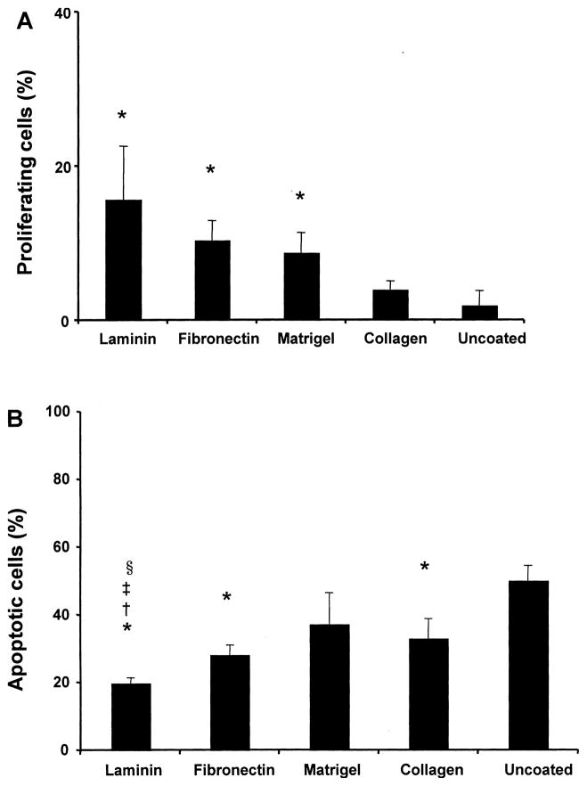
Figure 3.
Proliferation and apoptosis of statically seeded RAEC. (A) The percentage of proliferating RAEC after 24 h of culture on coated/uncoated scaffolds. Surface coating of PGS scaffold with laminin, fibronectin, and Matrigel resulted in significantly more proliferative endothelial cells (*p < 0.05 vs. uncoated scaffold, n = 3 slides counted, Ave. ± SD). (B) The percentage of apoptotic cells expressed as the proportion of TUNEL-positive cell nuclei to the total number of nuclei (*p < 0.01 vs. uncoated, ‡p < 0.05 vs. fibronectin, †p < 0.05 vs. Matrigel, §p < 0.05 vs. collagen, n = 3 slides counted, Ave. ± SD).

