
Figure 6.
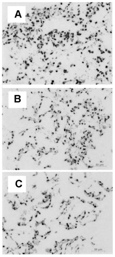
Representative micrographs of H&E staining after 72 h in the absence of flow (A), under low flow (B), or high flow (C). Cells remained well adhered and spread out in the PGS scaffolds even at the high flow rate.
Official websites use .gov
A
.gov website belongs to an official
government organization in the United States.
Secure .gov websites use HTTPS
A lock (
) or https:// means you've safely
connected to the .gov website. Share sensitive
information only on official, secure websites.
Representative micrographs of H&E staining after 72 h in the absence of flow (A), under low flow (B), or high flow (C). Cells remained well adhered and spread out in the PGS scaffolds even at the high flow rate.