Figure 3.


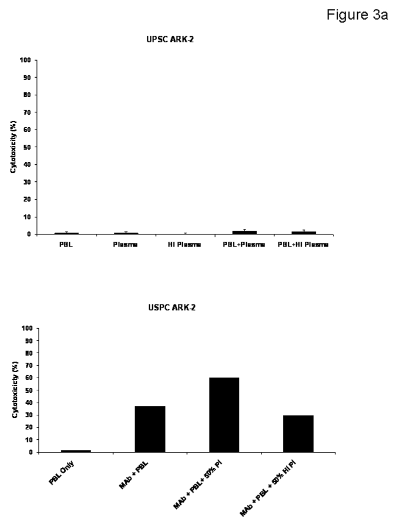
Figure 3a. USPC cell lines were challenged by adding human plasma diluted 1:2 (with or without heat inactivation) in the presence or absence of the effector cells and MT201 to standard 5-h 51Cr release assays. The addition of PBLs with or without plasma (untreated or heat-inactivated) was not able to induce significant cytotoxicity against USPC ARK-2 cell line (Upper Panel). Addition of physiological concentrations of IgG (i.e., heat-inactivated plasma diluted 1 : 2) to PBL in the presence of MT201 did not significantly alter the degree of ADCC achieved in the presence of MT201 (lower panel). In contrast, addition of untreated plasma (diluted 1:2) to PBL in the presence of MT201 consistently increased MT201-mediated cytotoxicity against USPC (p < 0.03) (lower panel).
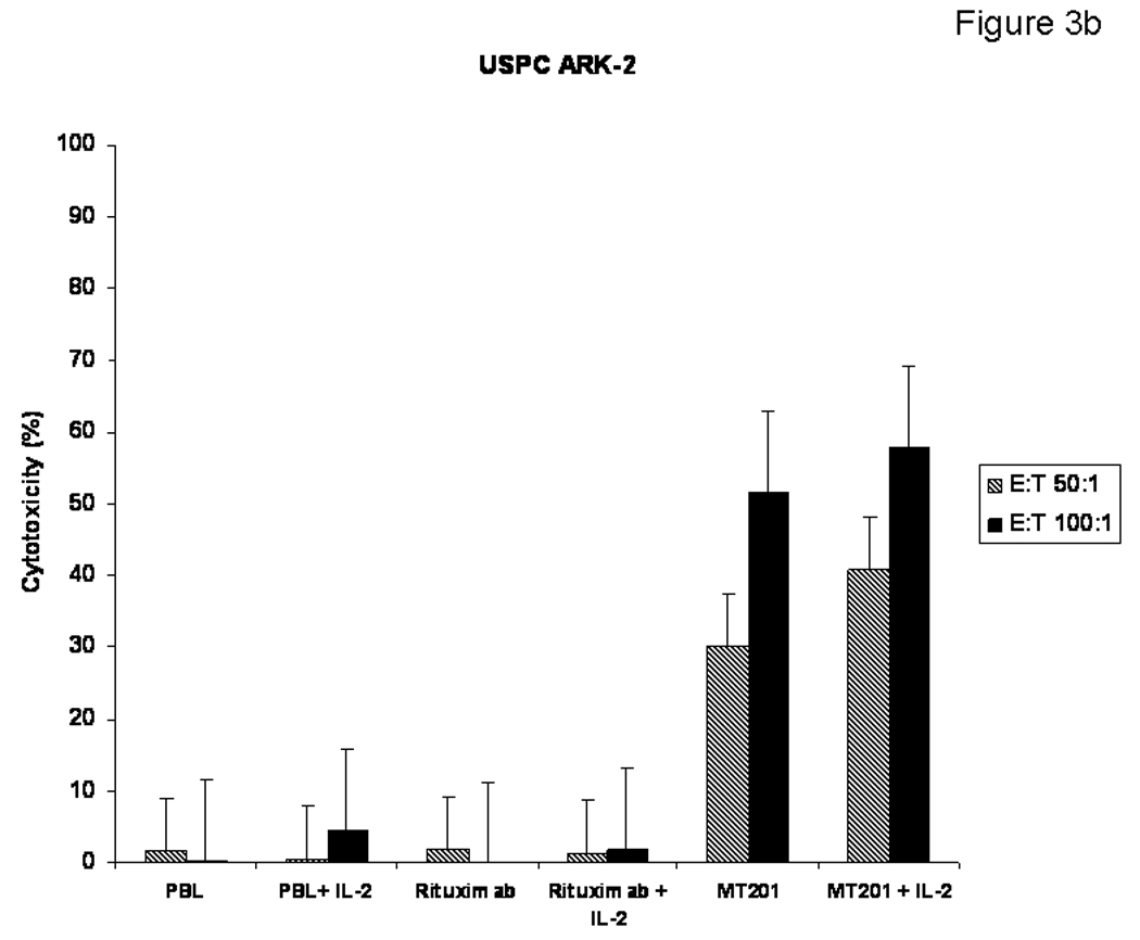
Figure 3b. The effect of low doses of interleukin-2 (IL-2) in combination with MT201 (5 µg/ml) on ADCC against USPC cell lines. PBL from healthy donors were incubated for 5 to 72 h in the presence of 100 IU/ml of IL-2. MT201-mediated ADCC was significantly increased in the presence of low doses of IL-2. No significant increase in cytotoxicity was detected after 5-h IL-2 treatment in the absence of MT201 or in the presence of rituximab isotype control mAb.
