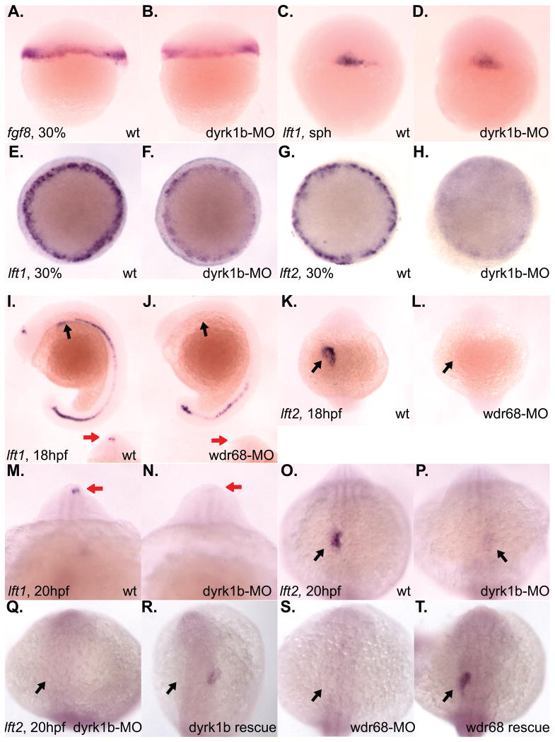
Figure 4. Reduced expression of lft1 and lft2 in dyrk1b and wdr68 knockdown animals.
A) lateral view of normal fgf8 expression in control animals at 30% epiboly stage. B) normal fgf8 expression in dyrk1b-MO animals at 30% epiboly stage. C) lateral view of normal lft1 expression in control animals at sphere (sph) stage. D) normal lft1 expression in dyrk1b-MO animals at sphere stage. E) dorsal view of normal lft1 expression in control animals at 30% epiboly stage. F) reduced lft1 expression in dyrk1b-MO animals at 30% epiboly stage. G) dorsal view of normal lft2 expression in control animals at 30% epiboly stage. H) reduced lft2 expression in dyrk1b-MO animals at 30% epiboly stage. I) lateral view of normal lft1 expression in control animals at 18hfp. Black arrows indicate asymmetric expression in the lateral plate mesoderm of the developing heart field. Red arrows in inset images are on the left side of the animals and indicate asymmetric expression in the diencephalon. J) severely reduced lft1 expression in the asymmetric heart and diencephalon territories of wdr68-MO animals at 18hpf. K) dorsal view of normal lft2 expression in control animals at 18hpf. Black arrows indicate asymmetric expression in the lateral plate mesoderm of the developing heart field. L) severely reduced lft2 expression in the asymmetric heart and diencephalon territories of wdr68-MO animals at 18hpf. M) anterior view of normal lft1 expression in the asymmetric diencephalon territory of control animals at 20hpf. Red arrows are on the left side of the animals and indicate asymmetric expression in the diencephalon. N) severely reduced lft1 expression in the asymmetric diencephalon territory of dyrk1b-MO animals at 20hpf. O) dorsal view of normal lft2 expression in the asymmetric heart territory of control animals at 20hpf. Black arrows indicate asymmetric expression in the lateral plate mesoderm of the developing heart field. P) severely reduced lft2 expression in the asymmetric heart territory of dyrk1b-MO animals at 20hpf. Q) dorsal view of severely reduced lft2 expression in dyrk1b-MO animals co-injected with EF1alpha transcripts. R) partial rescue of lft2 expression in dyrk1b-MO animals co-injected with Dyrk1b transcripts. S) dorsal view of severely reduced lft2 expression in wdr68-MO animals co-injected with EF1alpha transcripts. T) partial rescue of lft2 expression in wdr68-MO animals co-injected with FLAG-Wdr68 transcripts.

