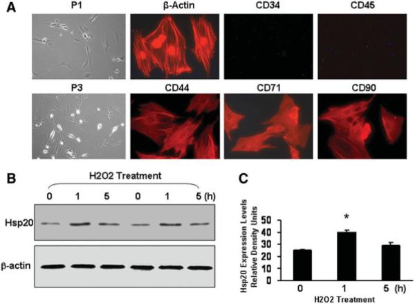
Figure 1.
Characterization of mesenchymal stem cells (MSCs) isolated from male rat bone marrow and time course of Hsp20 expression in response to H2O2 treatment (200 μM). (A): Cultured P3 MSCs were immunostained with primary antibodies as indicated. Alexa Fluor 594 goat anti-rabbit or mouse IgG was used as a secondary antibody and also negative controls. Images of phase contrast are at original magnification, ×100; the immunocytochemistry images are at original magnification, ×400. (B): At the indicated intervals, MSCs treated with H2O2 were lysed to assess Hsp20 expression by Western blot analysis. Panel (C) shows the quantitative results from the Western blots. Similar results were observed in three additional, independent experiments. β-Actin was used as internal control. Abbreviations: Hsp, heat-shock protein; P1, passage 1; P3, passage 3.

