
Figure 3.
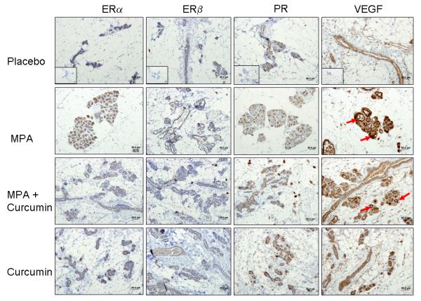
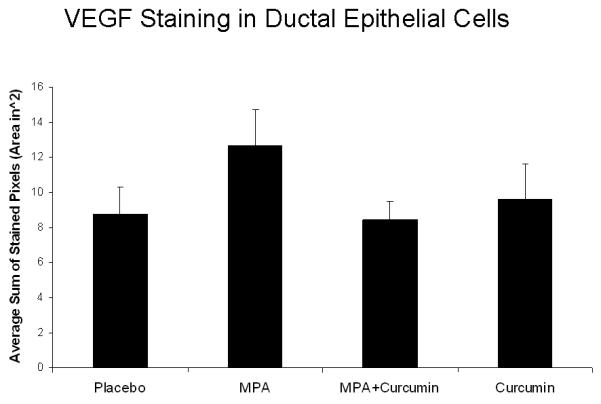
(A) ER and PR expression are unaffected by curcumin and VEGF levels are decreased by curcumin in MPA + curcumin treated DMBA-induced mammary tumors. Mammary gland tissues were collected at the conclusion of the study on day 52, sectioned, and immunostained for ER-α, ER-β, PR and VEGF as described in the Methods section. No significant differences were observed in the intensity of the staining among the treatment groups for ER-α, ER-β, and PR, however, curcumin blocked MPA-driven increases in VEGF levels in hyperplastic lesions (red arrows). Insets represent negative controls with no primary antibody staining for each antibody. (B) Photographs of slides were analyzed using FoveaoPro 3.0 analysis software. Positive VEGF staining was quantified as the number of VEGF-positive pixels in 3 different fields. Though not statistically different, the amount of VEGF was reduced in the group treated with MPA + Curcumin compared to the group treated with MPA alone. No significance was determined when analyzed using ANOVA.