Figure 6.
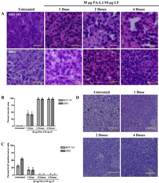
PA-L1/LF treatment exhibits dramatic anti-tumor activity in late stage disease. Mice bearing well-established BHT-101 or DRO tumors were treated with either 1, 2, or 4 doses of 30 μg PA-L1/10 μg LF. Animals receiving multiple doses were treated every other day. After the last scheduled dose, the animal was euthanized and tumor responses were compared to animals not receiving therapeutic intervention. H&E staining demonstrates extensive necrosis in both BHT-101 and DRO tumors (A). Scale = 20 μm, total magnification 400x. (B) BHT-101 (light gray bars) necrosis increased to 43.5% with a single dose of PA-L1/LF while DRO (dark gray bars) necrosis increased 41.5%. Both tumors exhibited ≥98% necrotic area with two doses of 30 μg PA-L1/10 μg LF. (C) BHT-101 (light gray bars) and DRO (dark gray bars) tumors exhibited a corresponding 13.9 and 37.9% decrease of Ki-67 expression, respectively. Two doses of 30 μg PA-L1/10 μg LF induced essentially 100% senescence of tumor cell proliferation and no significant change with 4 PA-L1/LF doses. (D) Representative DRO tumors from each dose number are shown. Error bars represent SEM. *unpaired t test p value <.05. Scale = 100 μm, total magnification 100x

